Electrocardiogram
What Is an Electrocardiogram? - By Dr. Kedar Kulkarni
Dr. Kedar Kulkarni, among the best cardiologists in Pune, told about the Electrocardiogram.
The electrocardiogram (ECG or EKG) is a diagnostic tool that is routinely used to assess the electrical and muscular functions of the heart. While it is a relatively simple test to perform, the interpretation of the ECG tracing requires significant amounts of training.
The heart is a two-stage electrical pump and the heart’s electrical activity can be measured by electrodes placed on the skin. The electrocardiogram can measure the rate and rhythm of the heartbeat, as well as provide indirect evidence of blood flow to the heart muscle.
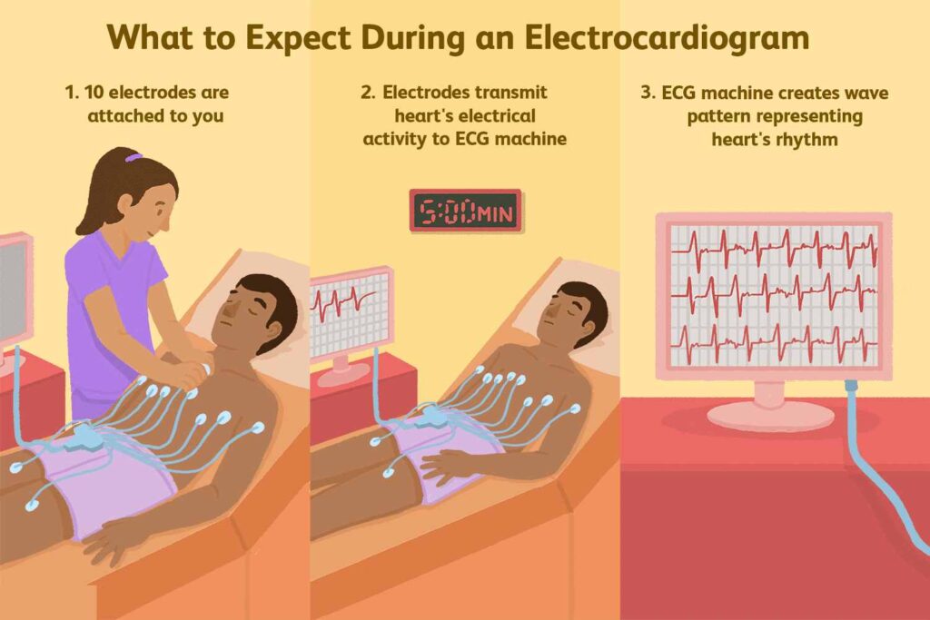
A standardized system has been developed for the electrode placement for a routine ECG. Ten electrodes are needed to produce 12 electrical views of the heart. An electrode lead, or patch, is placed on each arm and leg and six are placed across the chest wall. The signals received from each electrode are recorded. The printed view of these recordings is the electrocardiogram.
What Happens During an ECG? Is It Painful?
The ECG is a relatively simple test to perform. It is non-invasive and does not hurt. Patches are placed on the skin to detect electrical impulses that the heart generates. These impulses are recorded by an ECG machine. Four patches are placed on the limbs. One is placed on each shoulder or upper arm and one on each leg. These are called limb leads. There are six patches that are placed on the chest wall beginning just to the right of the breast bone. Patches are placed in the shape of a semi-circle ending near the left axilla (underarm). These are called the chest leads. These patches are connected to an ECG machine that records the tracings and prints them onto paper.
ECG Basics
ECG rate refers to how fast the heartbeats. Normally, the SA node generates an electrical impulse 50-100 times per minute. Bradycardia (brady=slow+cardia=heart) describes a heart rate of fewer than 50 beats per minute. Tachycardia (tachy=fast+cardia=heart) describes a heart rate faster than 100 beats per minute.
ECG Rhythm refers to the type of heartbeat. Normally, the heart beats in sinus rhythm with each electrical impulse generated by the SA node resulting in a ventricular contraction, or heartbeat. There are a variety of abnormal electrical rhythms, some are normal variants and some are potentially dangerous. Some electrical rhythms do not generate a heartbeat and are the cause of sudden death.
ECG Axis. If the QRS is upright (more positive than negative) in leads I and aVF, the axis is normal. The normal axis range is –30 degrees to +105 degrees.